Infraclavicular Brachial Plexus Block

Indications: Upper extremity surgical procedures below the level of the shoulder and deltoid.
Special Considerations: Particularly useful for bilateral upper extremity blockade, because phrenic nerve involvement is sparred. Ideal for very small children to decrease pneumothorax risk, as compared to the supraclavicular block location. Provides excellent coverage for hand and forearm surgery, that includes blockade of the area of tourniquet placement (interscalene does not provide full coverage of the hand / medial forearm, axillary does not provide coverage of a mid-humeral level tourniquet). Often considered a more challenging block, due to the deeper positioning of the sub-arterial target at this location.
Patient Position: Ideally, arm abducted laterally with elbow flexed, making a 90-degree angle, however, this block is useful in that it is possible to place with the arm at nearly any orientation. A folded towel or other padding placed beneath the shoulder can aid in bringing the deep artery to a more superficial position.
Dose: 0.5 - 1.5 mg/kg of Bupivacaine or Ropivacaine (roughly 0.2 - 0.4 mL/kg).
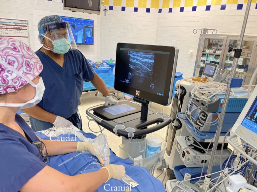
Technique: Probe – Linear; Needle – In-plane
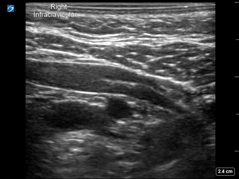
Placing the probe perpendicularly over the mid clavicle, the subclavian artery and vein are identified in cross section. The probe then is moved as far out laterally as visualization allows, past the lung apices, to the point where the subclavian artery just transitions into the axillary artery (as the subclavian artery crosses the lateral border of the first rib, it becomes the axillary artery). Needle entry is in-plane, from a cranial-to-caudal approach. Target location is 6 o’clock on the subclavian-axillary artery. The goal is deposition of local anesthetic in a u-shaped pattern surrounding the artery. Ideally, the artery will compress and lift with each incremental injection. If this pattern is seen, it is not necessary to redirect the needle to deposit addition local anesthetic at the 12 o’clock position on the artery.
Coverage: Brachial plexus, cord level.
Potential Complications:
Intravascular injection or injury
Pneumothorax (if needle directed too medially)
Bleeding / infection at needle insertion site
Patient Positioning and Probe Orientation
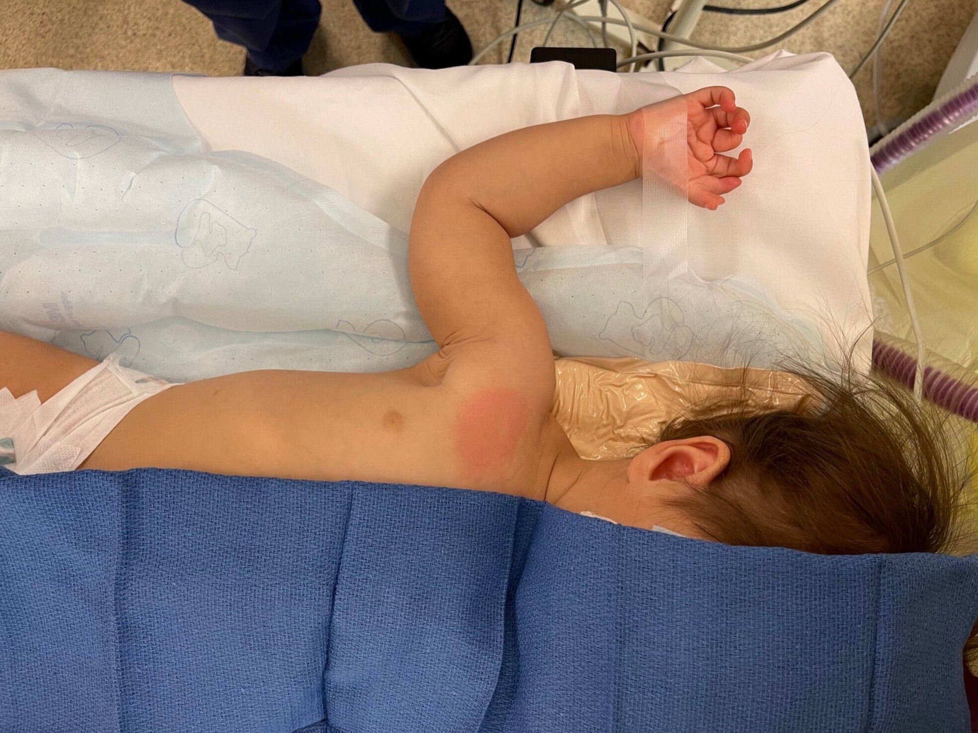

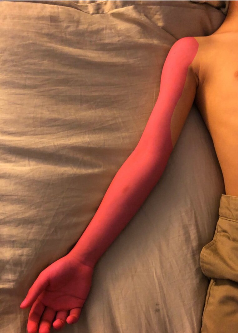
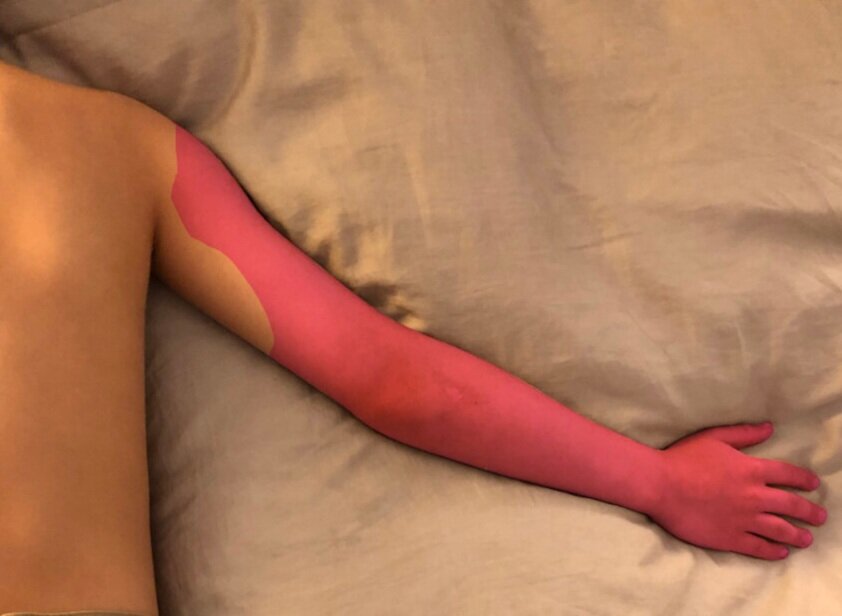
Expected Dermatomal Coverage
Ultrasound Images


Ultrasound guidance for pediatric infraclavicular nerve block was first described in 2004 by Marhofer, et al., who noted faster onset times and longer duration of sensory blockade as compared to nerve stimulation technique. Multiple approaches to this block – regarding patient positioning, ultrasound probe orientation, needle entry in relation to the clavicle, in-plane versus out-of-plane technique – are possible. Choosing the best technique will mainly depend on the approach which offers an individual provider the most comfort and success, but must also take into account patient comorbidities, and the surgical case at hand. Of note, one study (Yayik, et al.) compared the costoclavicular method to the lateral sagittal approach (described above) and found similar duration of anesthesia and post-operative pain scores, but faster block performance time.
Regarding concentration of local anesthetic for this block, the author’s preference is to use 0.2% Ropivacaine, to allow for differential sensory and motor blockade, with 1:200,000 epinephrine as an intravascular marker. If one’s goal is to achieve both a dense sensory and motor block, 0.5% Ropivacaine or 0.25-0.5% Bupivacaine would be appropriate options. If you or the surgeon are concerned about postoperative compartment syndrome, 0.125% ropivacaine offers adequate analgesia while allowing for the reassurance of pressure- and acidosis-derived pain to break through. Lower local anesthetic volumes are ideal for pediatric infraclavicular brachial plexus blockade. Similar sensory blockade and post-operative pain scores can be achieved with 0.25ml/kg as compared to 0.5ml/kg of local anesthetic volume (Ince, et al.). Because of the compact neurovascular bundle of the brachial plexus at this location, adequate coverage almost never requires more than 10-15mL of volume and can be achieved with far less in our smallest of patients.
In summary, the pediatric infraclavicular brachial plexus block offers a similar distribution of coverage as the supraclavicular block, but its lateral placement helps to decrease possible inadvertent puncture of the lung apices and avoids phrenic nerve involvement. Its relatively challenging depth can be navigated with ultrasound guidance and it offers the ability to avoid high doses of local anesthetic volume.
Ince I, Aksoy M, Dostbil A, Tuncer K. Can we use lower volume of local anesthetic for infraclavicular brachial plexus nerve block under ultrasound guidance in children? J Clin Anesth. 2017;41:132-136.
Marhofer P, Sitzwohl C, Greher M, Kapral S. Ultrasound guidance for infraclavicular brachial plexus anaesthesia in children. Anaesthesia. 2004;59(7):642-646.
Yayik AM, Cesur S, Ozturk F, Celik EC, Naldan ME, Ahiskalioglu A. Comparison of the lateral sagittal and costoclavicular approaches for ultrasound-guided infraclavicular block in pediatric patients: a prospective randomized study. Braz J Anesthesiol. Published online June 6, 2021:S0104-0014(21)00224-4.